1.1 The Skeletal System
1.1.1 Distinguish anatomically between the axial and appendicular skeleton.
1.1.2 Distinguish between the axial and appendicular skeleton in terms of function.
1.1.2 Distinguish between the axial and appendicular skeleton in terms of function.
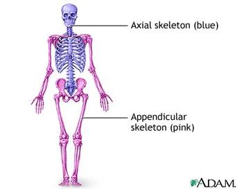
The axial skeleton is fixed - little movement occurs
The appendicular skeleton consists of the bones that are responsible for movement (includes the synovial joints)
- Skull
- Ribs
- Sternum
- Vertebral Column (cervical - 7 bones, thoracic - 12 bones, lumbar - 5 bones, sacral - 5 bones (fused as one), and coccyx - 4 bones (fused as one).
The appendicular skeleton consists of the bones that are responsible for movement (includes the synovial joints)
- Pectoral Girdle (scapulae and clavicles)
- Humurus
- Radius
- Ulna
- Carpals
- Metacarpals
- Phalanges
- Pelvic Girdle (Ilium, Ischium and Pubis)
- Femur
- Patella
- Tibia
- Fibula
- Tarsals
- Metatarsals
1.1.3 State the four types of bones
Ossification: the process of bone formation, the conversion of cartilage into bone
Long Bone
Short Bone
Flat Bone
Irregular Bone
Long Bone
- longer than they are wide
- works as levers (used with bones in the arms and legs)
- long cylindrical shaft, with enlarged ends
- essential for movement
Short Bone
- cube shaped bones
- small and compact
- provide strength for intricate movements
- carpal and tarsal bones
Flat Bone
- have curved surfaces
- vary from thick to thin
- protects organs
- allow muscle attachment
- skull, ribs, sternum and scapula
Irregular Bone
- all other bones that do not fall into the other categories
- they have varied size, shape and surface area
- include vertebral bones
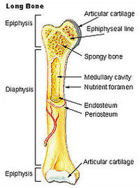
1.1.4 Draw and annotate the structure of a long bone
Need:
epiphysis,
spongy bone,
articular cartilage,
diaphysis,
compact bone (outer layer),
bone marrow,
marrow (medullary) cavity,
blood vessel (the red outer line) and
periosteum.
epiphysis,
spongy bone,
articular cartilage,
diaphysis,
compact bone (outer layer),
bone marrow,
marrow (medullary) cavity,
blood vessel (the red outer line) and
periosteum.
1.1.5 Apply anatomical terminology to the location of bones
- Anterior, front side of the body, also known as ventral.
- Posterior, back side of the body, also known as the dorsal.
- Distal, farthest end from the trunk or head.
- Proximal, closest part nearest the trunk or head.
- Inferior, below also, toward the feet.
- Superior, above or near the head, also known as cranial.
- Lateral, away from the midline.
- Medial, toward the midline.
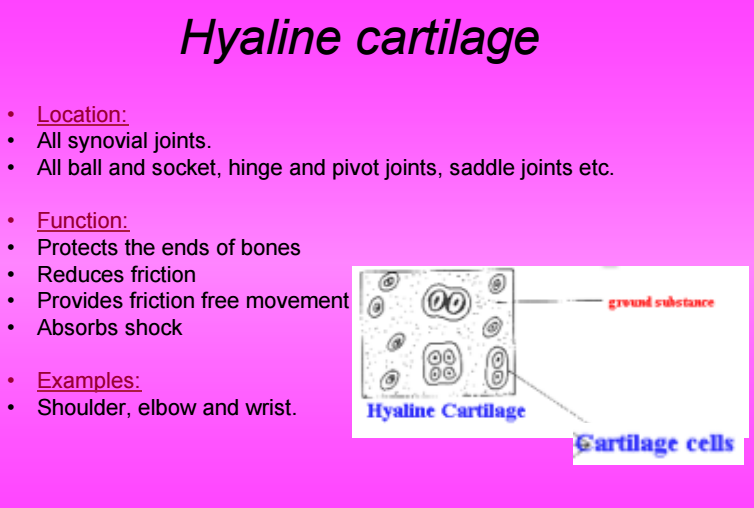
1.1.6 Outline the functions of connective tissues
|
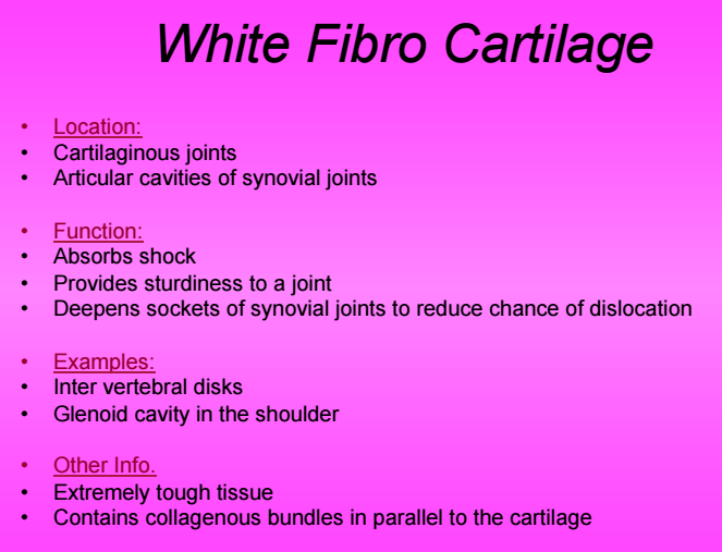
Cartilage
|
|

1.1.7 Define the term joint
"A joint occurs where two or more bones articulate"
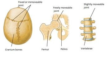
1.1.8 Distinguish between the different types of joint in relation to movement permitted
|
Fixed/Immovable joints
Cartilaginous/slightly moveable joints
Synovial/freely movable joints
|
|
|
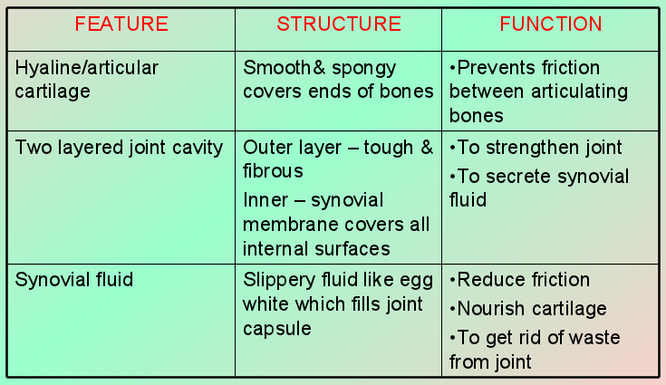
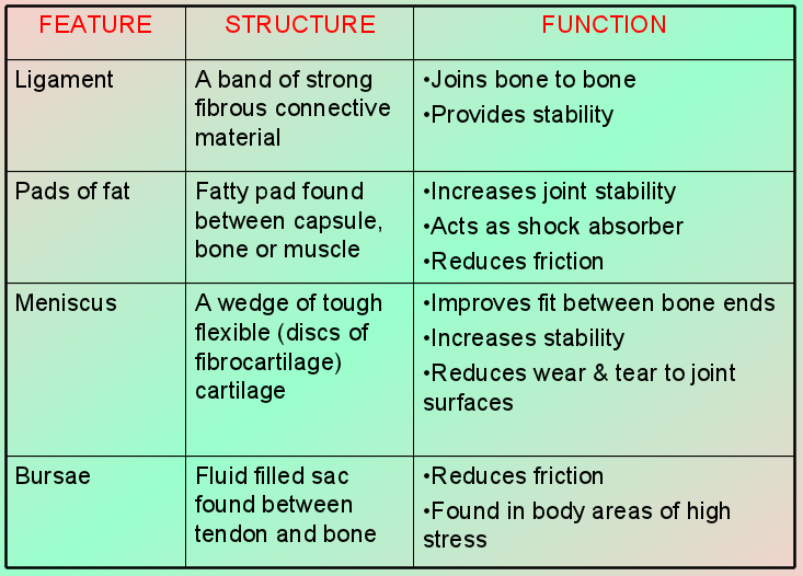
1.1.9 Outline the features of a synovial joint
Articular cartilage, synovial membrane, synovial fluid, bursae, miniscus, ligaments, and articular capsule |
|
|
1.1.10 List the different types of synovial joints
|
Joint
Gliding Joint between the tarsal bones and carpal bones Hinge Joint elbow or knee joint Pivot Joint Radioulnar Joint Bottom of the skull Condyloid Joint Wrist joint Saddle Joint The thumb Ball and Socket Joint Shoulder joint Hip joint |
Description
Usually flat or slightly curved bones,, slide across each other Least amount of movement of all the synovial joints. The articular surfaces have been fused together so that movement is only permitted in one direction The articular surfaces are joined by strong ligaments Movement is only allowed in one plane (extension / flexion) Rounded surface of one bone rolls around a ring formed by bone and ligament A ball shaped bone fits into a cup Saddle shaped bone fits into a bone shaped like the legs can move up and down and side to side A sphere shaped bone fits into the rounded cavity of the other covered in cartilage to prevent friction High range of movement |
Sporting Example
Used for bending of the wrist in a baseball hit Extending the leg when striking a football to score a goal Extending the arm when making a hit in boxing Heading a football Serving in tennis where the wrist is primarily used Gripping a tennis racket Movement coming from the hip in kicking a football |
1.2 The Muscular System
1.2.1 Outline the general characteristics common to muscle tissue
Contractility - ability of muscle to shorten
Extensibility - ability of muscle to lengthen
Elasticity - ability of muscle to return to normal size
Atrophy - the wasting away of muscle tissue
Hypertrophy - the increase in size of muscle tissue
Controlled by nerve stimuli - requires an impulse or recognition to initiate movement
Fed by capillaries - blood will be redistributed to muscles when they are working and require more oxygen
1.2.2 Distinguish between the different types of muscle
Skeletal - Under voluntary control, has a striated appearance. Has tendons that attach mostly to bone. Main function of this type of muscle is to move the skeleton
Cardiac - Also known as heart muscle. Also striated but under involuntary control. Contracts without you having to think about it
Smooth - Lines the walls of blood vessels and hollow organs such as the stomach and intestines. Also involuntary.
Cardiac - Also known as heart muscle. Also striated but under involuntary control. Contracts without you having to think about it
Smooth - Lines the walls of blood vessels and hollow organs such as the stomach and intestines. Also involuntary.
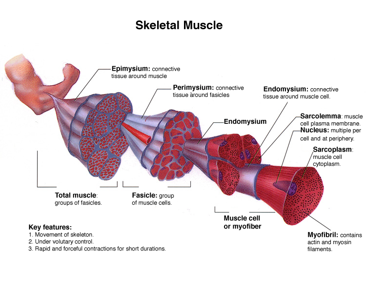
1.2.3 Annotate the structure of skeletal muscle
1.2.4 Define the terms origin and insertion of muscles
origin: Origin is the attachment of a muscle tendon to a stationary bone
insertion: Insertion is the attachment of a muscle tendon to a moveable bone
origin: Origin is the attachment of a muscle tendon to a stationary bone
insertion: Insertion is the attachment of a muscle tendon to a moveable bone
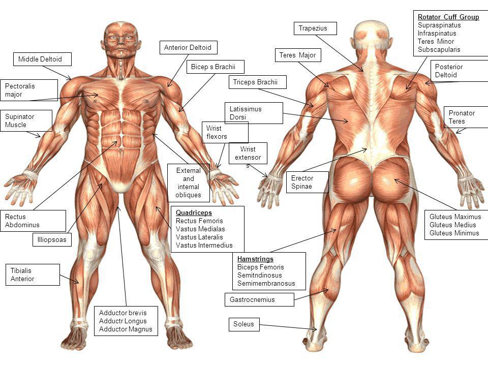
1.2.5 Identify the location of skeletal muscles in various regions of the body